How Color Blindness Works: Cones, Rods & CVD Explained
2026-03-18 · Jamie Chen · 11 min read
TL;DR: Cones encode color; this guide explains CVD types—then take the free 18-plate screen.
You’re looking at a stoplight. The person next to you sees it the same way you do - or do they? About one in twelve men and roughly one in two hundred women experience color vision deficiency, meaning their eyes process color information differently than the majority of people. But here’s what most people don’t realize: this isn’t a flaw. It’s a variation in how the biology of the eye works.
If you’ve ever wondered why some people struggle to distinguish red from green, or why certain colors seem to blend together, the answer lies deep inside your retina, in millions of specialized cells called cones and rods. Understanding how these cells function - and what happens when they don’t - is the key to understanding color blindness itself.
How the eye sees color
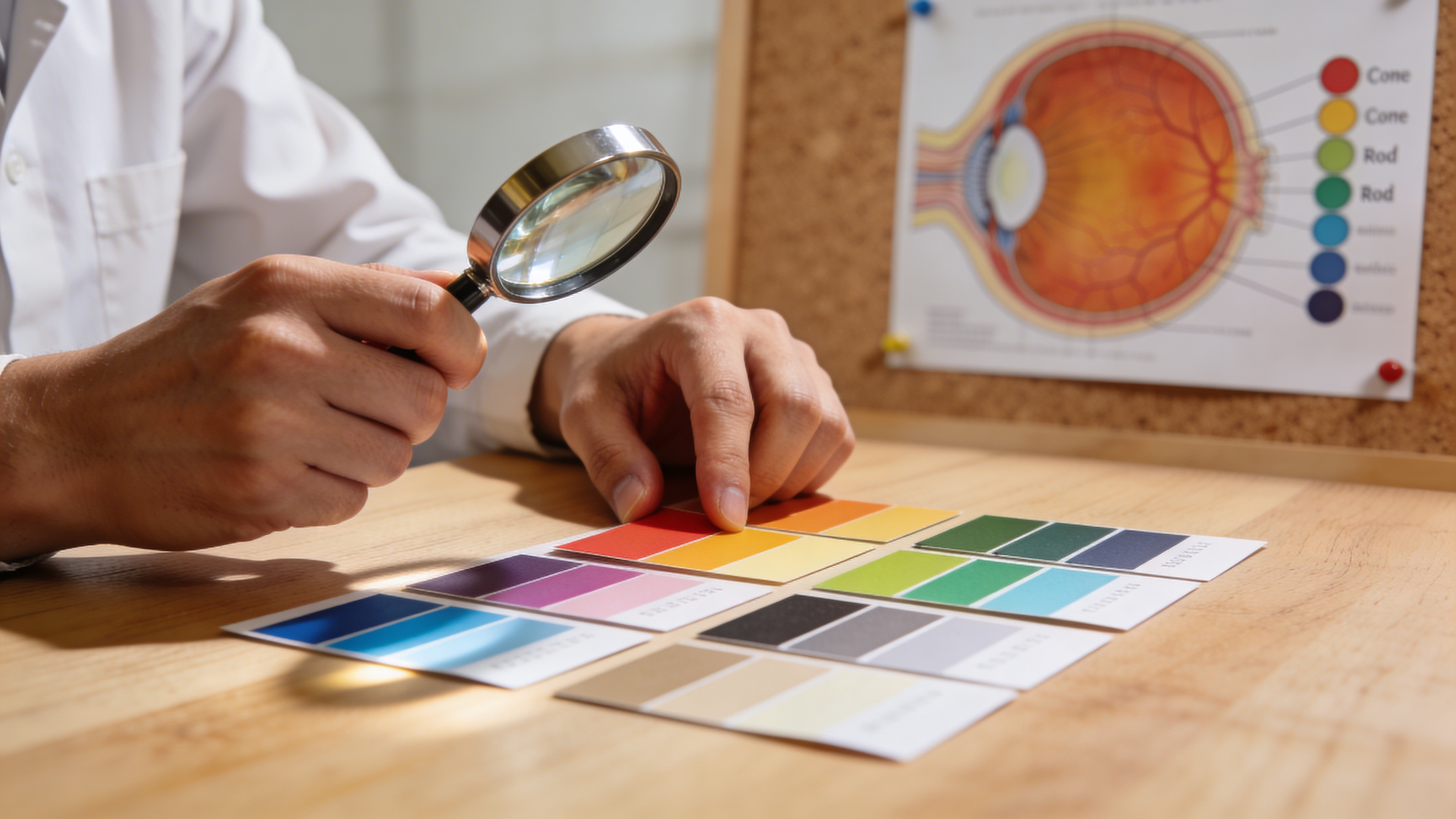
Let’s start with the basics. When light enters your eye, it travels through the cornea and lens, which focus it onto the back of your eye called the retina. The retina is a thin layer of tissue about the thickness of a postage stamp, and it’s packed with light-sensitive cells. Two types of cells do this work: rods and cones.
Rods are incredibly sensitive to light. They handle your vision in low-light conditions - like reading a book at dusk or navigating a dark room. But rods can’t distinguish color. They see in grayscale. That’s why everything looks gray and colorless when the lights go down.
Cones are different. They function best in bright light and they’re responsible for color vision. Each cone cell contains light-sensitive proteins called photopigments. These pigments absorb photons of light at specific wavelengths, and that’s what creates the sensation of color in your brain.
There are three types of cones, and each one is tuned to a different part of the light spectrum. This is where color vision gets interesting.
The three types of cones and how they create color
The human eye has roughly 6 to 7 million cone cells. About 64 percent of them are sensitive to longer wavelengths of light - what we call red light. These are called L-cones (long-wavelength cones). About 32 percent are sensitive to medium wavelengths, what we perceive as green light. These are M-cones (medium-wavelength cones). Only about 2 percent are sensitive to short wavelengths, or blue light. These are S-cones (short-wavelength cones).
Here’s the crucial part: color isn’t a property of light itself. It’s something your brain constructs by comparing the signals from all three cone types. When you look at an orange, light of multiple wavelengths bounces off it and enters your eye. The L-cones fire heavily, the M-cones fire moderately, and the S-cones fire less. Your brain takes this pattern of signals and assembles it into the experience of “orange.”
When you see yellow, the L-cones and M-cones are both firing strongly, while S-cones barely respond. Your brain interprets this particular pattern as yellow. Purple? That’s L-cones and S-cones firing together while M-cones stay quiet. Every color you’ve ever seen is your brain’s interpretation of the ratio of signals coming from these three cone types.
This is why color is, in a real sense, a collaboration between your eye and your brain. Two people with identical eyes might still experience color slightly differently based on individual differences in how their brains process those signals.
What happens in color vision deficiency
Color blindness isn’t actually blindness. The term is misleading. Most people with CVD can see color just fine. What they have is a difference in how their cones work.
The most common form of color vision deficiency is red-green color blindness, which accounts for roughly 99 percent of all CVD cases. This happens when either the L-cones or the M-cones - or both - don’t function properly.
Protanopia (red blindness) occurs when L-cones are missing or non-functional. People with protanopia have only two working cone types: M-cones and S-cones. Without red-sensitive cones, they lose an entire channel of color information. Colors that depend on red input - like pure red, orange, and many shades of brown - become invisible or shift into yellows and browns. A red apple might look brown or dark yellow. Traffic lights are a common challenge: the red light becomes indistinguishable from the dark background, while the green and yellow lights remain visible (though yellow-green, which normally combines red and green signals, might look off).
Deuteranopia (green blindness) is when M-cones are absent or don’t work. People with deuteranopia still have L-cones and S-cones. The loss of green sensitivity means colors that rely on green signals shift. Red and green appear more similar to each other. A red stoplight might appear yellow or beige. Grass looks more yellow-brown. Like protanopia, it’s red-green confusion, but the specific way colors shift is different.
Tritanopia (blue blindness) is much rarer, affecting roughly one in fifty thousand people. It occurs when S-cones are absent or malfunction. People with tritanopia retain L-cones and M-cones but lose blue sensitivity. This means blues and yellows become hard to distinguish. The sky might look pink or white. Yellows might look pink. It’s a different kind of color confusion entirely, and it presents different challenges in everyday life.
Achromatopsia (complete color blindness) is the rarest form. It occurs when all three cone types fail to function, or when the cones themselves are absent. People with achromatopsia see the world in shades of gray, similar to a black-and-white photograph. But unlike the black-and-white films you’ve seen, their world is often very sensitive to light and can be visually uncomfortable in bright conditions. They may also experience nystagmus, an involuntary eye movement that can affect focus.
The genetics behind color vision deficiency
Why do some people have non-functional cones while others don’t? The answer is genetics.
The genes that code for L-cones and M-cones are located on the X chromosome. Women have two X chromosomes, so they have two copies of these genes. Men have only one X chromosome, so they have just one copy. This is why color blindness is more common in men: if their single X chromosome carries a gene for CVD, they’ll express it. Women would need a CVD gene on both X chromosomes - much rarer.
That said, women can be carriers of red-green color blindness. A carrier woman has one normal gene and one CVD gene. She typically has normal color vision because her normal gene is usually enough. But she can pass the CVD gene to her children.
There’s another possibility: women can develop color vision deficiency through a condition called X-inactivation, where one of their X chromosomes is randomly “turned off” in many of her cells. If the X chromosome carrying the normal gene gets inactivated in a significant portion of her cone cells, she might experience some degree of red-green color blindness. This is rare but it happens.
The gene for tritanopia is located on chromosome 9, not the X chromosome, so it affects men and women roughly equally. Achromatopsia involves mutations in genes related to cone function more broadly and can be inherited in various patterns.
Severity and variation
Here’s something important: color vision deficiency isn’t binary. It’s not simply “you have it” or “you don’t.”
Some people have what’s called anomalous trichromacy, where all three cone types are present but one of them is shifted - it’s sensitive to wavelengths slightly different from the standard range. This usually causes mild color confusion. The person might struggle with specific color pairs but see most colors normally. Red-green confusion might only show up when the red and green are very similar in hue.
Others have dichromacy, where one cone type is completely absent or non-functional. These are the cases typically called protanopia, deuteranopia, and tritanopia.
The severity also depends on which cones are affected and to what degree. Some people might have L-cones that are only partially functional, creating a milder version of protanopia. Others might have cones that are completely absent, creating a more severe form.
This is why color blindness exists on a spectrum. Two people both diagnosed with protanopia might experience color quite differently depending on the exact nature of their cone malfunction.
Acquired color vision deficiency
It’s worth noting that color blindness isn’t always present from birth. Some people develop CVD later in life due to disease or injury.
Diabetes, glaucoma, and age-related macular degeneration can all affect cone function. Certain medications can also cause acquired color blindness. Vitamin B12 deficiency can impact color perception. Eye trauma or stroke can damage the parts of the brain responsible for processing color signals.
This is one reason why a sudden change in your color perception is worth discussing with an eye care professional. While congenital color blindness (present from birth) is stable and won’t worsen, acquired color blindness can sometimes be managed or arrested if caught early.
Why this matters in daily life
Understanding the biology of color vision deficiency goes beyond academic interest. It has real consequences for how we design the world.
If you know that protanopia involves the loss of red information, you understand why using red and green as the only distinction in a visualization fails for about one percent of the population. You understand why a traffic signal needs to rely on position or shape, not just color. You understand why color alone should never be the only way to communicate information in a graph, interface, or map.
For people with CVD, understanding the science behind their own color vision can be empowering. It’s not a defect. It’s a variation in cone function. Some research even suggests that people with certain types of color blindness might have advantages in specific visual tasks - for example, detecting camouflaged objects that blend into a red-green background.
Diagnosis and screening
If you’re curious about your own color vision, several screening methods exist. The most famous is the Ishihara test, which uses colored dot patterns to detect red-green color blindness. There are also more sophisticated tests like the Farnsworth-Munsell 100 Hue test, which asks you to arrange colored caps in order. An eye care professional can also perform specialized tests using color matching or chromatic discrimination. You can start with a quick online color vision test before booking a formal exam.
Many cases of color vision deficiency go undiagnosed. People adapt without realizing they see color differently. They might avoid certain careers or assume they’re just “bad with colors.” A screening can provide clarity.
When to talk to a professional
If you notice a sudden change in your color perception - colors that used to seem normal now look off, or you’re struggling with color distinctions you never struggled with before - schedule an appointment with an ophthalmologist or optometrist. Acquired color blindness can signal an underlying health issue that deserves attention.
If you have a family history of color vision deficiency and you’re planning to pursue a career where color discrimination matters (like aviation, certain trades, or certain medical fields), it’s worth getting screened early. Some professions have specific requirements about color vision.
If you’re a parent and you think your child might have color blindness, early screening can help. It won’t change the biology, but it can help you and your child’s teachers understand their needs and adapt accordingly.
The future of understanding color vision
Research into color vision deficiency continues. Scientists are exploring the possibility of gene therapy to restore or modify cone function. Some early trials have shown promise, though this technology is still in experimental stages and not yet available as a standard treatment.
There’s also growing recognition that designing for people with CVD isn’t about accommodating a disability - it’s about designing better for everyone. High-contrast designs, non-color indicators, and thoughtful color choices benefit people with and without color vision deficiency.
FAQ
Can color blindness be cured? Congenital color blindness (present from birth) cannot currently be cured. It’s a genetic trait, not a disease. However, early-stage gene therapy research shows promise for the future. Acquired color blindness (from disease or injury) might sometimes be managed or arrested if the underlying cause is treated, so it’s worth discussing any sudden changes with a professional.
Is color blindness always inherited? Congenital color blindness is inherited through genes. Red-green color blindness is typically passed down through the X chromosome. However, acquired color blindness can develop at any time due to disease, medication, or injury - these cases are not inherited.
Can women be color blind? Yes, though it’s rarer. Women need a CVD gene on both X chromosomes, which is uncommon. However, some women can be carriers and pass the gene to their children. Additionally, conditions like X-inactivation can sometimes allow a carrier woman to experience color blindness. Women can also develop acquired color blindness.
Do people with color blindness see everything in black and white? Only people with achromatopsia (complete color blindness) see in black and white. People with red-green color blindness (the most common form) see colors - just different colors than people with typical vision. Someone with protanopia might see blues and yellows clearly but struggle to distinguish reds and greens.
Is color blindness the same as being colorblind? The terms are used interchangeably, but “color vision deficiency” is more medically precise. “Color blindness” is simpler but technically misleading since most people with CVD can see color. In this article, both terms refer to the same condition.
The science of color vision is elegant and complex. Your cones and rods are doing an incredible job every moment you see. When they work slightly differently - when one type of cone isn’t there or doesn’t function as expected - the experience of color shifts. Understanding that shift is the first step toward designing a world where everyone, regardless of how their cones work, can navigate and enjoy it equally.
Want to understand how you or someone else perceives color? Take our free Ishihara-style color blind test to get instant results, then explore real-world examples in the color blindness simulator.
Related Articles
- Practical accessibility tools overview
- How to use a color blindness simulator for images and websites
- Simulation methodology (Machado 2009)
Get a clear picture of your own color vision with DeficiencyView’s free color blind test - Ishihara-style results in under two minutes, no login required.